Drew Ludwig · 7 October 2026
Laboratory Recreations of Opal Formation Processes Yield Insights into Silica Nanostructure Evolution
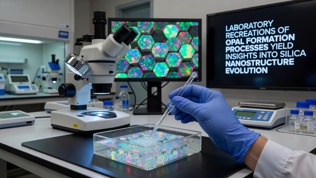
Researchers in materials science have turned to controlled laboratory environments to replicate the slow geological processes that create natural opals, and these experiments continue to reveal details about how silica nanoparticles assemble into ordered structures. The work focuses on the self-assembly of silica spheres that form the photonic lattices responsible for opal's characteristic play of color, and it draws from both mineralogy and colloid chemistry to map out the steps involved. Natural opals develop when silica dissolves from surrounding rocks, travels through groundwater, and deposits into cavities where spheres pack together over long periods. Laboratory teams accelerate this sequence by using sol-gel techniques and controlled precipitation, which allows them to adjust sphere size, uniformity, and packing density in weeks rather than millennia. Data from these setups show that even small variations in pH or temperature during synthesis alter the final lattice quality and therefore the optical properties observed in the resulting material.Methods Used in Controlled Silica Sphere Synthesis
Teams typically start with tetraethyl orthosilicate as a precursor, hydrolyzing it under acidic or basic conditions to generate monodisperse silica nanoparticles ranging from 150 to 300 nanometers in diameter. Once formed, the spheres settle in a solvent and arrange themselves through evaporation or sedimentation, creating face-centered cubic lattices that diffract visible light. Observers note that maintaining narrow size distributions remains critical, because deviations larger than five percent disrupt the ordered packing and reduce color intensity.
Additional variables tracked during these runs include ionic strength of the solution and the rate of solvent removal, both of which influence whether defects such as stacking faults or vacancies appear in the final array. Researchers at institutions in Australia and Germany have published protocols that combine microfluidics with these traditional methods, enabling real-time monitoring of assembly dynamics through optical microscopy and small-angle X-ray scattering.
Structural Evolution Observed During Assembly
As spheres pack, they first form disordered aggregates that gradually reorganize into crystalline domains, a transition documented through time-resolved imaging. The process reveals intermediate stages where grain boundaries migrate and anneal, ultimately producing larger single-crystal regions that exhibit stronger Bragg diffraction. Measurements collected in October 2026 at facilities equipped with synchrotron sources confirmed that annealing at moderate temperatures after initial deposition improves long-range order without altering sphere diameter.
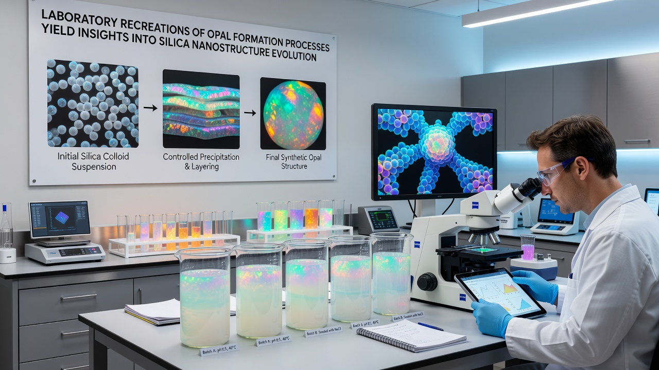
Further analysis using electron microscopy and spectroscopy shows how point defects and line defects propagate through the lattice and how these imperfections scatter light at wavelengths outside the main photonic band gap. Such findings help explain why some synthetic opals display muted colors compared with high-quality natural specimens mined in regions like Lightning Ridge.
Applications and Ongoing Research Directions
Insights gained from these laboratory models extend beyond gemology into the design of photonic crystals and sensors. Because the same assembly principles govern both natural and synthetic systems, engineers adapt the protocols to create films and coatings that filter specific wavelengths or respond to environmental changes. A 2025 report from the Commonwealth Scientific and Industrial Research Organisation outlines how scaled-up versions of these processes could support optical computing components, while parallel work at the Massachusetts Institute of Technology explores inverse opals as catalyst supports.
Cross-disciplinary teams continue to refine simulation tools that predict lattice formation from initial particle interactions, reducing the need for trial-and-error experimentation. These computational models incorporate hydrodynamic forces and van der Waals attractions, yielding results that align closely with experimental outcomes recorded under varying humidity and pressure conditions.
Conclusion
Laboratory recreations of opal formation therefore serve as a practical bridge between geological observation and nanoscale engineering, supplying quantitative data on how silica nanostructures evolve from dispersed particles into functional photonic materials. Continued refinement of synthesis parameters and characterization techniques promises additional clarity on defect control and optical performance, supporting both scientific understanding and technological development in adjacent fields.